MICROSCOPY / INSTRUMENTATION
Super-cool microscopes
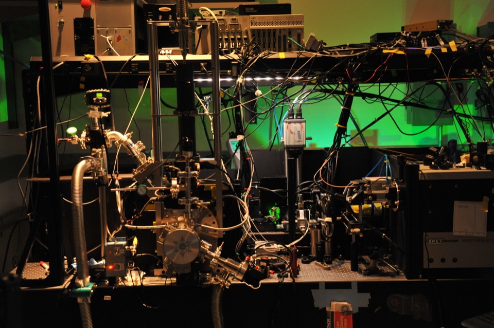
Our correlative pipeline is now available at the AIC @ Janelia
OPTICS · MICROSCOPY · CODE
My name is David P. Hoffman. I’m a scientist/engineer who builds robust, high-performance optical systems and loves data and Python
I’ve probed ultrafast chemical reactions, imaged nanoscale protein ultrastructure relationships, and implemented a high throughput screening platform. Now I’m applying optics to omics.
Drag to explore. Zoom with + and −, then pinch or drag on touch. Arrow keys pan; 0 resets.
01 / ABOUT

I’m a scientist and engineer with a proven track record of hardware and software development whose research has been featured on the cover of Science magazine.
In graduate school at Berkeley I trained as a physical chemist and spectroscopist before diving into imaging and biology as a post-doc in Nobel Laureate Eric Betzig’s group.
While I enjoy digging into the nuts and bolts of instrumentation, theory and code, I always see my work in the context of the big picture and strive to do the most useful thing most efficiently. I’m goal focused, data driven and led by curiosity.
Interests: Microscopy 🔬 · Lasers ⚡️ · Python 🐍 · My daughter’s Legos 🧱
Find me on GitHub ↗02 / SELECTED WORK
MICROSCOPY / INSTRUMENTATION
Our correlative pipeline is now available at the AIC @ Janelia
LASERS / SPECTROSCOPY
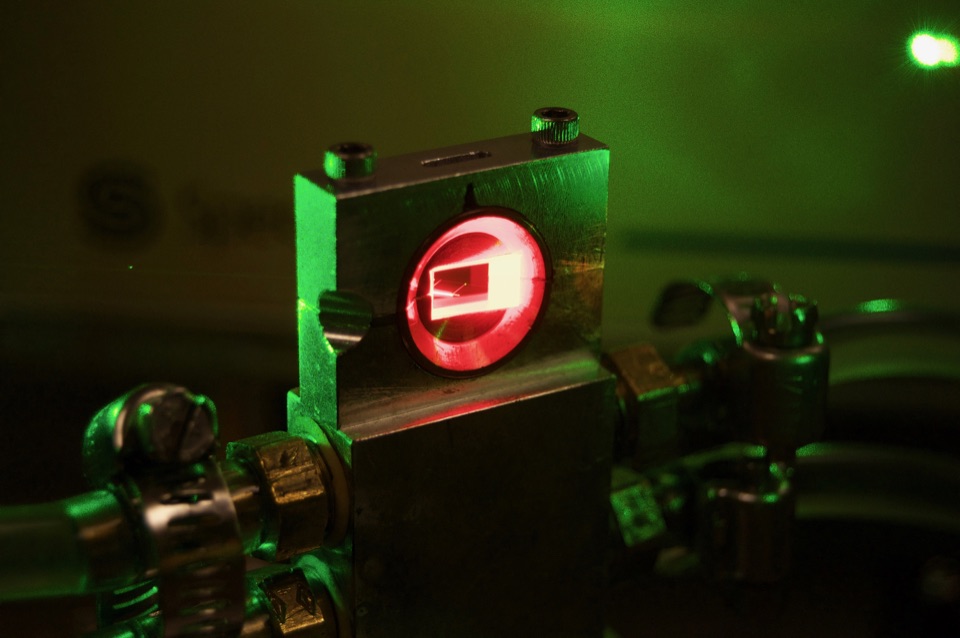
In graduate school I used femtosecond spectroscopy to interogate molecular reactions
Read the thesis ↗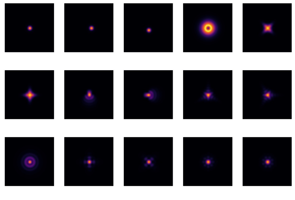
PYTHON / COMPUTATIONAL IMAGING
A Python package for simulating optical transfer functions and point spread functions for light microscopes
Explore pyOTF ↗More optics + omics: Xenium
April 2022 – Present
Optics + omics
April 2020 – March 2022
I was developing the company’s core hardware technology platform, a high throughput single particle tracking (SPT) microscope. My focus was increasing throughput as efficiently as possible by engineering advanced illumination and analysis strategies. As a small startup I wore many other hats such as: developing our data analysis pipeline, introducing the use of Git and GitHub, helping set up company operations, and designing the company logo.
October 2019 – April 2020
HHMI, Janelia Research Campus ↗
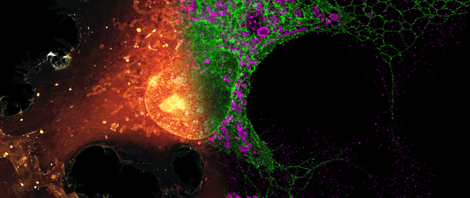
I developed an advanced pipeline to collect and correlate data taken from a cryogenic super resolution fluorescent microscope (which I designed and built) and a customized focused ion beam scanning electron microscope (FIB-SEM), to elucidate key questions in cell biology, research which was featured on the January 17th, 2020 cover of Science magazine.
August 2014 – October 2019
University of California, Berkeley ↗
I spent my time in graduate school applying femtosecond stimulated Raman spectroscopy (FSRS) to multiple chemical systems. In particular, I developed 2D-FSRS which offers an unprecedented view of chemical reaction dynamics. During my tenure, I rebuilt many of the optical subsystems of the spectrometer, such as the Ti:Sapphire femtosecond oscillator and the non-colinear optical parametric amplifier (NOPA), vastly improving system performance. In addition, I rewrote the analysis software and rewrote the LabVIEW instrument control software. In total, the changes I made increased mean time to failure from ~2 hours to months.
September 2008 – May 2014
EDUCATION & TRAINING
04 / WRITING
lengthy, confused, and inconsequential
Seriously, don’t do it, it’s not helpful
Getting really good real resolution in SMLM is really hard.
How to make your images look bad with high NA objectives
The back pupil plane of a microscope objective is Fourier conjugate to it’s image plane. We use this fact to perform laser scanning microscopy or create the interference patterns necessary for SIM or LLSM.
My basic modus operandi has been to learn and then, if not immediately, soon after forget them. This is a list I maintain of things once a remember them.
05 / PUBLICATIONS
Correlative three-dimensional super-resolution and block-face electron microscopy of whole vitreously frozen cells
Cells need to compartmentalize thousands of distinct proteins, but the nanoscale spatial relationship of many proteins to overall intracellular ultrastructure remains poorly understood. Correlated light and electron microscopy approaches can help. Hoffman et al. combined cryogenic super-resolution fluorescence microscopy and focused ion beam–milling scanning electron microscopy to visualize protein-ultrastructure relationships in three dimensions across whole cells. The fusion of the two imaging modalities enabled identification and three-dimensional segmentation of morphologically complex structures within the crowded intracellular environment. The researchers observed unexpected relationships within a variety of cell types, including a web-like protein adhesion network between juxtaposed cerebellar granule neurons.
Science · Read the paper ↗What role do vibrational coherences, if any, play in condensed phase photoreactivity? While we have observed vibrational coherences in almost any system where we look hard enough, it hasn’t been clear if these are a cause or consequence of reactivity. In this survey of our own work we argue that for fast reactions (< 10 ps) coordinate, coherent nuclear motion is required.
Accounts of Chemical Research · Read the paper ↗06 / GET IN TOUCH